Anatomical Urinary System Model Of Male Internal Reproductive Disease
Approx $83.38 USD
Introduction to the Anatomical Urinary System Model of Male with Internal Reproductive Disease
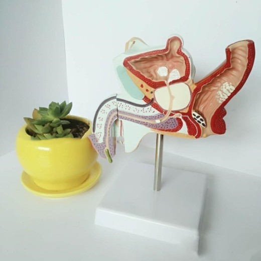
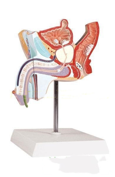
The Anatomical Urinary System Model of the Male with Internal Reproductive Disease is a detailed educational tool designed to demonstrate male urinary and reproductive anatomy alongside common diseases affecting these systems. Featuring the kidneys, bladder, prostate, testes, and relevant vascular structures, this model is ideal for studying conditions such as prostate disease, bladder disorders, and reproductive health issues. Suitable for medical students, healthcare professionals, and educators, this model provides a hands-on way to explore urinary and reproductive anatomy, making it a valuable resource in anatomy classrooms, medical training centers, and urology clinics across New Zealand.
H2: Key Features of the Anatomical Urinary System Model of Male with Internal Reproductive Disease
1. Realistic Depiction of Male Urinary and Reproductive Anatomy
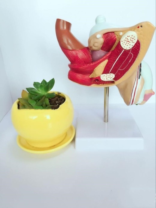
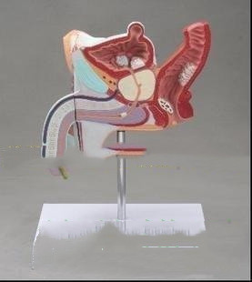
The model provides a realistic and comprehensive view of male urinary and reproductive anatomy, including structures such as the kidneys, ureters, bladder, prostate gland, seminal vesicles, testes, and vas deferens. Each component is crafted to accurately reflect its structure and function, allowing users to explore the interconnectedness of these systems and understand how they work together to manage urinary function and reproductive health.
2. Illustration of Common Diseases and Pathologies
This model highlights common male urinary and reproductive diseases, such as prostate enlargement, urinary tract infections, and testicular lesions. These conditions are depicted to demonstrate the physical changes and disruptions caused by disease, providing insight into their effects on urinary flow, reproductive health, and organ function. This feature is particularly useful for understanding how specific pathologies impact overall health and well-being.
3. Cross-Sectional Views for In-Depth Study
The model includes cross-sectional views of key organs, such as the bladder and prostate, allowing users to observe internal structures and understand how diseases develop within these systems. Cross-sectional views help to clarify the spatial relationships and pathways within the urinary and reproductive tracts, supporting deeper learning about disease mechanisms and their physiological impact.
4. Color-Coded and Labeled Structures for Easy Identification
To assist in identifying each structure, the model includes color-coded sections and clear labels for major organs and tissues. This color-coding enhances understanding by distinguishing the kidneys, ureters, bladder, prostate, and reproductive structures, making it easier to recognize normal anatomy and pathological changes. This feature is especially beneficial for visual learners and medical students new to the study of urology and reproductive health.
5. High-Quality and Durable Construction
Made from premium, non-toxic materials, this model is designed for durability and frequent handling, making it ideal for educational and clinical use. Its high-quality build ensures that it retains detail and integrity over time, even with regular use in classrooms and clinical settings in New Zealand. This durable construction makes the model a reliable resource for long-term study and patient education.
6. Compact and Display-Ready Design
The model is designed to be portable and compact, allowing for easy placement on desks, tables, or shelves. Its stable base keeps it secure during study sessions or consultations, making it ideal for anatomy classrooms, urology clinics, and healthcare training centers. The display-ready design makes it convenient for a range of educational and clinical settings, enhancing its versatility as a teaching tool.
H2: Why Choose the Anatomical Urinary System Model of Male with Internal Reproductive Disease?
1. Essential for Medical and Urology Education
This model is essential for medical students and healthcare professionals learning about the male urinary and reproductive systems, providing a realistic view of these complex anatomical structures and common disease states. For New Zealand’s healthcare trainees and urology students, this model bridges theoretical knowledge and practical understanding, helping to illustrate the physical manifestations of disease in the urinary and reproductive organs.
2. Ideal for Patient Education and Urological Consultations
In clinical settings, this model is invaluable for explaining conditions related to male urinary and reproductive health to patients. By visually demonstrating disease progression and physical changes, healthcare providers can help patients better understand diagnoses and treatment options. This model aids in communication and promotes informed decision-making for patients concerned with urological or reproductive issues.
3. Supports Visual and Kinesthetic Learning
The model caters to both visual and kinesthetic learners, offering a hands-on experience that enhances comprehension and retention. Color-coded structures support visual learning, while detailed anatomical features allow kinesthetic learners to interact directly with the model, making complex anatomy and disease pathology more accessible.
4. Useful for Exam Preparation and Clinical Assessments
For students preparing for exams or clinical assessments, the model serves as a valuable study aid. Its realistic design and labeled structures make it easy to review anatomy and disease processes, reinforcing essential knowledge for exams in anatomy, physiology, and urology. Medical students and healthcare trainees benefit from this hands-on approach to studying urinary and reproductive health, enhancing retention and confidence.
5. Educational Display for Clinics and Classrooms
Beyond its educational value, the Anatomical Urinary System Model serves as an informative display for clinics, classrooms, and healthcare facilities. In clinical environments, it provides a visual aid for explaining urinary and reproductive health, helping to promote awareness of common conditions and preventative care. For New Zealand educators and healthcare providers, this model supports a well-rounded understanding of male health.
H2: Maintenance and Care Tips for Your Anatomical Urinary System Model of Male with Internal Reproductive Disease
To keep your model in excellent condition, follow these care tips:
-
Dust Regularly: Use a soft cloth or brush to remove dust from the model, particularly around intricate areas. Regular
cleaning preserves the model’s appearance and prevents dirt buildup.
-
Avoid Direct Sunlight: Display the model away from direct sunlight to prevent colors from fading over time. Indoor display
helps maintain its quality and vibrant look.
-
Handle with Care: Although durable, handle the model gently to avoid damaging smaller parts or delicate areas, especially
if it includes detailed depictions of lesions or other pathology.
- Store Properly When Not in Use: When not displayed, store the model in its original packaging or a dust-free area to protect it from potential damage. Proper storage helps extend the model’s lifespan for long-term educational use.
Overview:
The model shows the male reproductive qi forest disease, urethral knots, benign prostatic hyperplasia, sharp warts, spermatogonia liu and
other diseases and their pathological manifestations. It adopts a one-to-one ratio design and selects the anatomical area of the male
reproductive system.
Specification:
Size: 25.5X18X15CM
Type: Male internal reproductive pathology model
Style: School supplies
material: Plastic
Applicable people: General
Scope of application: General


The product may be provided by a different brand of comparable quality.
The actual product may vary slightly from the image shown.


.webp)
.webp)







.jpg)


.jpg)


.jpeg)


.jpeg)


.jpeg)


.jpeg)


.jpeg)

.jpeg)

.jpeg)

.jpeg)


.jpeg)
.jpg)

.jpeg)


.jpeg)
.jpeg)


.jpeg)


.jpeg)


.jpeg)

.jpeg)

.jpeg)

.jpeg)


.jpeg)
.jpeg)
.jpeg)


.jpeg)


.jpeg)


.jpg)
.jpeg)


.jpg)


ulva-Logo.jpg)


.jpeg)


.png)


.png)


