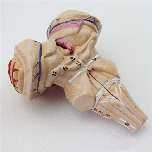
Brain Model 9 Parts With Artery Anatomical Model
Approx $77.41 USD
Introduction to the Brain Model: 9 Parts with Artery Anatomical Model
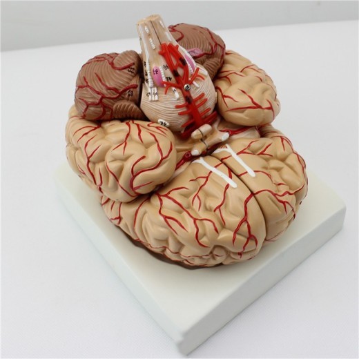
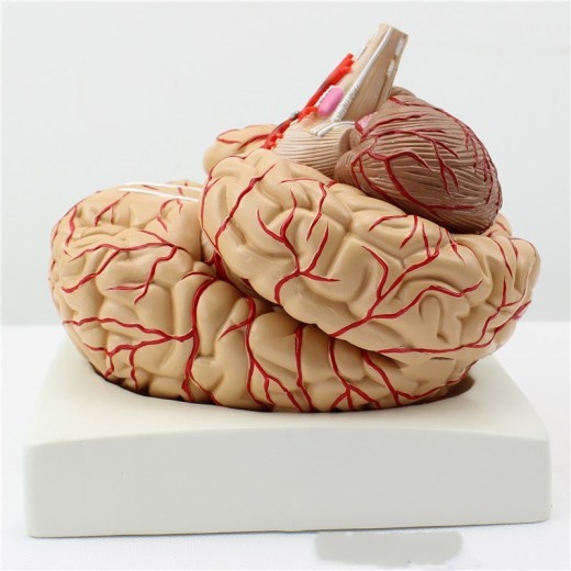
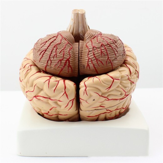
The 9-Part Brain Model with Arteries is a highly detailed educational tool designed to provide a thorough understanding of brain anatomy, including its arterial blood supply. This model, split into nine distinct sections, includes the major regions of the brain and critical arteries, making it ideal for studying neural structures, brain function, and cerebral circulation. Suitable for students, healthcare professionals, and educators, this brain model offers a hands-on approach to exploring brain anatomy, providing an invaluable resource for classrooms, medical training facilities, and clinics across New Zealand.
H2: Key Features of the Brain Model: 9 Parts with Artery Anatomical Model
1. Realistic 9-Part Segmentation of the Brain
The model is divided into nine detailed parts, including the frontal lobe, parietal lobe, temporal lobe, occipital lobe, cerebellum, brainstem, and more. Each part is crafted to reflect the structure and texture of the human brain accurately, allowing users to study the brain's complex organization and understand the function of each region. The segmentation provides a deeper understanding of how the brain’s regions interact and supports advanced studies in neurology and anatomy.
2. Detailed Arterial Representation
This model includes a detailed depiction of the brain’s arterial system, showcasing major arteries such as the anterior, middle, and posterior cerebral arteries, as well as the basilar and vertebral arteries. These arteries are essential for understanding the brain’s blood supply and provide insight into how oxygen and nutrients reach each part of the brain. The inclusion of the arterial system makes this model especially valuable for studying stroke mechanisms, blood flow, and other vascular-related brain functions.
3. High-Quality, Durable Construction
Constructed from premium, non-toxic materials, this model is designed to withstand regular handling, making it suitable for educational and clinical settings. Its durable build ensures that it remains intact even with frequent use, providing a reliable and long-lasting educational resource. This quality makes it ideal for anatomy classrooms, neurology labs, and healthcare training facilities in New Zealand.
4. Color-Coded Sections for Easy Identification
The Brain Model features color-coded sections that make it easier to distinguish between different regions and arteries. Each brain region and artery is marked with a distinct color to support quick identification and memorization of the brain’s structure. This color-coding system is particularly useful for visual learners, enhancing comprehension and retention of complex anatomical details.
5. Removable Parts for Hands-On Learning
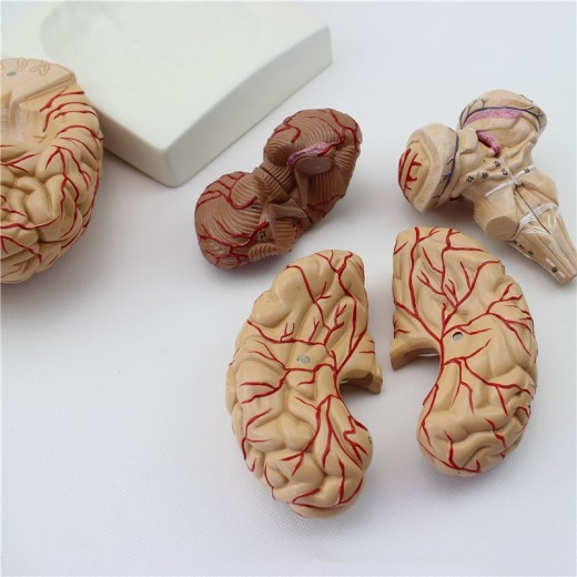
The nine parts of the brain are removable, allowing users to examine each region individually and study the brain’s inner structures. This feature makes the model ideal for advanced anatomy courses, where students can explore neural pathways, interconnections, and functional areas in more depth. By interacting with each component, learners gain a tangible understanding of brain anatomy that supports both theoretical and practical learning.
6. Compact and Display-Ready Design
With its compact and display-friendly design, this brain model can be placed on desks, shelves, or demonstration areas without taking up too much space. It includes a stable base to keep it secure during study sessions or demonstrations, allowing it to be easily moved between classrooms or consultation rooms. This versatility makes it an ideal addition to a range of educational and clinical settings.
H2: Why Choose the Brain Model: 9 Parts with Artery Anatomical Model?
1. Essential for Neurology and Health Education
This 9-part brain model is an essential tool for teaching brain anatomy, particularly for students in neurology, psychology, and health sciences. The detailed representation of brain structures and arteries provides a visual and hands-on experience that enhances understanding of brain function and organization. It helps bridge the gap between theoretical knowledge and practical application, making it a valuable resource for anatomy and medical education.
2. Ideal for Neurology, Psychology, and Neurosurgery Training
The Brain Model with Arteries is invaluable in training programs focused on neurology, psychology, and neurosurgery. Trainers can use it to illustrate critical concepts such as cerebral blood flow, brain function, and stroke mechanisms. For New Zealand’s healthcare training programs, this model provides an interactive approach to studying the brain, helping learners understand both structure and vascular function in detail.
3. Supports Visual and Kinesthetic Learning
The model is designed to cater to visual and kinesthetic learners, making it a versatile tool for different learning styles. The color-coded regions enhance visual learning, while the removable parts allow kinesthetic learners to explore each structure directly. This interactive approach enhances knowledge retention, making learning more effective and engaging.
4. Ideal for Exam Preparation and Clinical Assessments
For students preparing for exams or practical assessments, this model serves as a valuable study aid. Its realistic design and labeled components make it easy to review brain anatomy and blood supply, reinforcing critical knowledge needed for exams. Medical and neurology students can use this model to build confidence in identifying brain structures and understanding arterial pathways before assessments.
5. Educational Display for Clinics and Classrooms
Beyond its educational function, the Brain Model with Arteries serves as an informative display piece for clinics, classrooms, and neurology labs. In clinical settings, it can be used to explain brain-related conditions, such as stroke or aneurysms, to patients. This model is an effective tool for educators and healthcare providers in New Zealand to enhance awareness and understanding of brain anatomy, circulation, and health.
H2: Maintenance and Care Tips for Your Brain Model: 9 Parts with Artery Anatomical Model
To keep your Brain Model in excellent condition, follow these care tips:
-
Dust Regularly: Use a soft cloth or brush to dust the model, especially around the intricate areas and arteries. Regular
cleaning keeps the model looking sharp and prevents dirt buildup.
-
Avoid Direct Sunlight: Display the model away from direct sunlight to prevent fading of colors over time, maintaining its
vibrant appearance and quality.
-
Handle with Care: Although durable, handle the removable parts gently to avoid damaging delicate sections. Avoid forcing
parts together or apart to ensure the model remains functional and intact.
- Store Properly When Not in Use: When not on display, store the model in its original packaging or a protective case to prevent dust accumulation and potential damage. Proper storage helps extend the model’s lifespan for long-term educational use.
Product information:
Product category: emergency care
Scope of application: teaching demonstration
material: plastic
Weight: 900g
Product features: natural brain model, 9 parts, showing the basic morphological structure of the brain, and with arteries.
Color: brown
Size Information:
Size: 20*17*15cm


The product may be provided by a different brand of comparable quality.
The actual product may vary slightly from the image shown.

.webp)
.webp)





.jpg)


.jpg)


.jpeg)


.jpeg)


.jpeg)


.jpeg)


.jpeg)

.jpeg)

.jpeg)

.jpeg)


.jpeg)
.jpg)

.jpeg)


.jpeg)
.jpeg)


.jpeg)


.jpeg)


.jpeg)

.jpeg)

.jpeg)

.jpeg)


.jpeg)
.jpeg)
.jpeg)


.jpeg)


.jpeg)


.jpg)
.jpeg)


.jpg)


ulva-Logo.jpg)


.jpeg)


.png)


.png)


