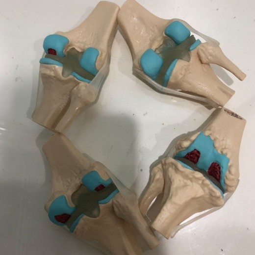
Four-Stage Diseased Knee Joint Model Sick Joint Model
Approx $53.21 USD
Introduction to the Four-Stage Diseased Knee Joint Model
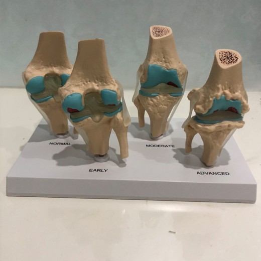
The Four-Stage Diseased Knee Joint Model is a valuable educational tool designed to demonstrate the progression of knee joint diseases, such as osteoarthritis, cartilage degeneration, and joint inflammation. This model depicts the knee joint in four distinct stages of disease, allowing students, healthcare professionals, and educators to observe how knee health deteriorates over time. By illustrating the effects of joint diseases on bone, cartilage, and ligaments, this model offers a hands-on way to study knee anatomy and pathology, making it an excellent resource for classrooms, clinics, and physical therapy centers in New Zealand.
H2: Key Features of the Four-Stage Diseased Knee Joint Model
1. Realistic Depiction of Knee Joint Degeneration
This model provides an accurate and realistic view of knee joint anatomy, showcasing changes in the bones, cartilage, and ligaments as the disease progresses. Each stage visually represents different levels of degeneration, helping users understand how conditions like osteoarthritis gradually affect joint structure. This realism is essential for understanding how knee diseases impact mobility and joint function.
2. Four-Stage Disease Progression
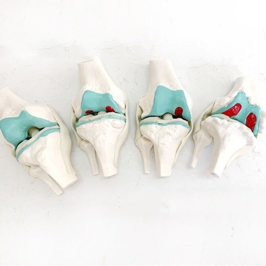
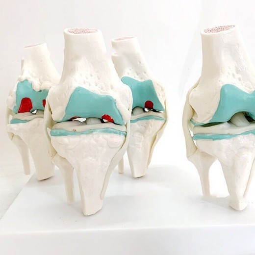
The model features four distinct stages of knee disease, starting with a healthy joint and progressing through mild, moderate, and severe degeneration. Each stage highlights specific changes, such as cartilage thinning, bone spurs, and increased inflammation. This progression helps students and professionals identify the characteristics of each stage, supporting learning about diagnosis, treatment, and patient care strategies.
3. Durable and High-Quality Construction
Made from durable, non-toxic materials, the Four-Stage Diseased Knee Joint Model is built for regular handling in educational and clinical settings. Its robust construction ensures that it remains intact even with frequent use, making it ideal for anatomy classrooms, orthopedic clinics, and physical therapy facilities. The high-quality materials provide a reliable resource for long-term study and demonstration in New Zealand.
4. Color-Coded and Labeled Parts for Easy Identification
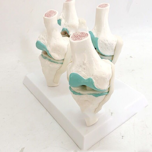
To enhance learning, the model includes color-coded sections to differentiate between healthy and diseased tissue, cartilage, and bone. The color-coded regions help users quickly identify degenerative changes and understand how the disease affects different parts of the knee joint. This feature supports visual learning and makes it easier to comprehend the progression of joint disease.
5. Cross-Sectional Views for In-Depth Study
The model often includes cross-sectional views that show the internal structure of the knee, making it easier to observe cartilage wear, bone deterioration, and joint space reduction. This cross-sectional design provides a closer look at the internal changes that occur as the disease advances, helping learners understand the underlying causes of pain, stiffness, and limited mobility.
6. Compact and Display-Ready Design
The compact design of the model makes it suitable for display on desks, shelves, or medical demonstration tables. Its stable base ensures that it remains secure during demonstrations and discussions, allowing for easy use in various educational and clinical settings. This display-ready feature makes it ideal for classrooms, orthopedic clinics, and physical therapy centers across New Zealand.
H2: Why Choose the Four-Stage Diseased Knee Joint Model?
1. Essential for Orthopedic and Physical Therapy Education
The Four-Stage Diseased Knee Joint Model is a critical tool for teaching joint anatomy and disease progression in orthopedic and physical therapy programs. It offers a tangible way to understand knee degeneration, providing insight into conditions such as osteoarthritis and cartilage wear. For students in New Zealand, this model bridges the gap between theory and practice, enhancing understanding of joint disease and patient care.
2. Ideal for Patient Education and Treatment Planning
This model is invaluable for patient education, helping healthcare providers explain knee conditions, treatment options, and potential outcomes. By visually demonstrating joint disease progression, the model allows patients to see how their knee health may change over time and understand the importance of preventative care or treatment. This visual aid supports effective communication and informed decision-making in clinical settings.
3. Supports Visual and Kinesthetic Learning
The model caters to both visual and kinesthetic learners, offering a hands-on approach that enhances engagement and comprehension. The color-coded parts support visual learning by distinguishing healthy tissue from diseased tissue, while the four stages allow kinesthetic learners to interact directly with the model. This interactive approach makes learning about knee health and disease progression more effective and memorable.
4. Valuable for Exam Preparation and Practical Assessments
For students preparing for exams or practical assessments, the Four-Stage Diseased Knee Joint Model serves as a valuable study aid. Its realistic design and labeled parts make it easy to review key anatomical changes associated with joint degeneration, reinforcing knowledge essential for exams. Medical, physical therapy, and orthopedic students benefit from this hands-on approach to studying joint pathology.
5. Educational Display for Clinics and Classrooms
Beyond its educational function, this model serves as an informative display in clinics, classrooms, and physical therapy facilities. In clinical settings, it can be used to educate patients on joint disease and treatment options, enhancing their understanding of joint health. For educators and healthcare providers in New Zealand, this model promotes awareness and understanding of knee diseases, supporting conversations about joint health and preventative care.
H2: Maintenance and Care Tips for Your Four-Stage Diseased Knee Joint Model
To keep your model in top condition, follow these care tips:
-
Dust Regularly: Use a soft cloth or brush to remove dust from the model, especially around color-coded sections and
intricate areas. Regular cleaning helps maintain the model’s appearance and prevents dirt buildup.
-
Avoid Direct Sunlight: Display the model away from direct sunlight to prevent colors from fading over time. Indoor
placement helps preserve the model’s quality and vibrant appearance.
-
Handle with Care: Although durable, handle the model gently to avoid damaging delicate parts, especially around areas
representing cartilage and bone structures.
- Store Properly When Not in Use: When not displayed, store the model in its original packaging or a dust-free area to protect it from potential damage. Proper storage helps extend the model’s lifespan for long-term educational use.
Product information:
Material: PVC
Size Information:
Specification: 28*17*16cm


The product may be provided by a different brand of comparable quality.
The actual product may vary slightly from the image shown.




.webp)
.webp)
.webp)
.webp)





.jpg)


.jpg)


.jpeg)


.jpeg)


.jpeg)


.jpeg)


.jpeg)

.jpeg)

.jpeg)

.jpeg)


.jpeg)
.jpg)

.jpeg)


.jpeg)
.jpeg)


.jpeg)


.jpeg)


.jpeg)

.jpeg)

.jpeg)

.jpeg)


.jpeg)
.jpeg)
.jpeg)


.jpeg)


.jpeg)


.jpg)
.jpeg)


.jpg)


ulva-Logo.jpg)


.jpeg)


.png)


.png)


