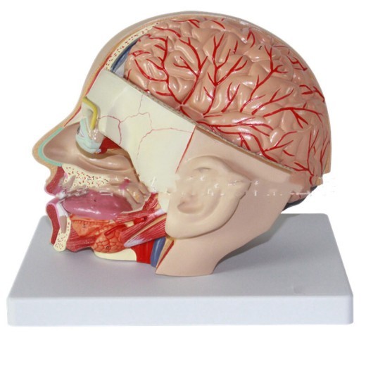
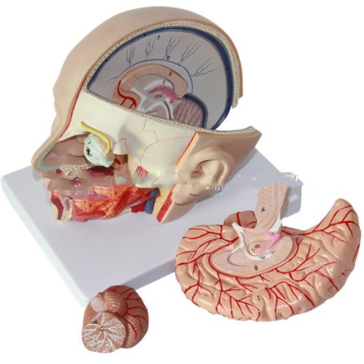
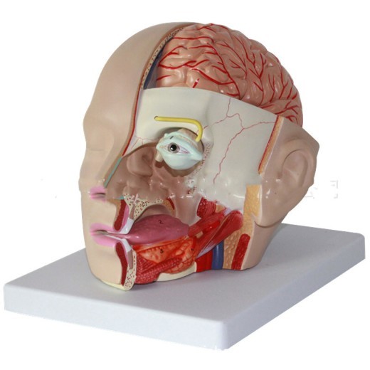
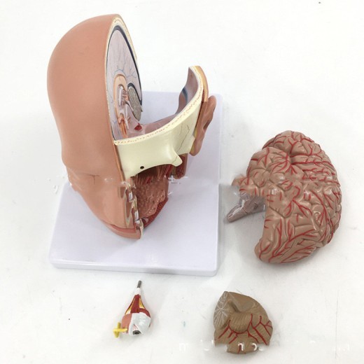
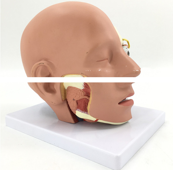
Human head anatomical model with brain model
Approx $142.59 USD
Introduction to the Human Head Anatomical Model with Brain Model
The Human Head Anatomical Model with Brain Model offers a comprehensive look at the anatomy of the human head, including detailed representations of the brain, skull, muscles, nerves, and other critical structures. This model provides an in-depth view of the head and brain, allowing students, educators, and healthcare professionals to study head anatomy and neural functions more effectively. Ideal for use in classrooms, medical schools, and healthcare training facilities across New Zealand, this model provides a three-dimensional, hands-on learning experience that enhances understanding of head and brain anatomy, making it an essential tool in medical education and patient education.
H2: Key Features of the Human Head Anatomical Model with Brain Model
1. Realistic and Detailed Head and Brain Anatomy
The Human Head Anatomical Model features an accurate depiction of key head structures, including the skull, facial muscles, nerves, blood vessels, and sinuses. The included brain model provides a comprehensive view of brain anatomy, highlighting areas such as the cerebrum, cerebellum, and brainstem. Each component is designed to reflect real anatomy, allowing users to study the connections and interactions between the head’s structures and the brain’s regions.
2. High-Quality, Durable Construction
Made from high-grade, non-toxic materials, this model is designed to withstand regular handling, making it ideal for classroom and clinical settings. Its durable construction ensures long-term use without compromising quality, providing a reliable educational resource for anatomy classrooms, medical labs, and healthcare training programs. This model is built to support years of study and demonstration, making it an excellent investment for New Zealand’s medical education institutions.
3. Color-Coded and Labeled Parts for Easy Identification
The model includes color-coded and labeled sections to help users easily identify critical structures, such as the skull bones, brain regions, cranial nerves, and blood vessels. Color-coding makes it simple to distinguish between parts, while labels provide clear guidance for identifying each structure. This feature is especially beneficial for visual learners, making complex anatomy easier to understand and remember.
4. Removable Brain Sections for In-Depth Study
The brain within the model is divided into multiple sections, including the frontal, parietal, temporal, and occipital lobes, as well as the cerebellum and brainstem. These sections can often be removed, allowing users to examine each part individually, providing a closer look at neural pathways and brain structures. This removable feature is especially valuable for advanced anatomy studies, giving students a tangible way to explore how the brain's different regions connect and interact.
5. Cross-Sectional View of the Head for Detailed Examination
The head model is designed to offer a cross-sectional view, displaying the internal structures of the head, such as the nasal cavity, oral cavity, and the cranial cavity that houses the brain. This cross-sectional feature enables users to understand the spatial relationships between the brain, sinuses, and other head components. This is particularly useful in ENT (Ear, Nose, Throat) education, neurology, and other medical disciplines where a complete understanding of head anatomy is essential.
6. Compact, Display-Ready Design
The model’s compact and stable design makes it ideal for display in a variety of settings, from classrooms and clinics to training centers. It is mounted on a sturdy base that keeps it stable during use, allowing for easy transport between teaching and consultation areas. This display-ready feature makes it a versatile tool, fitting seamlessly into different educational or clinical environments.
H2: Why Choose the Human Head Anatomical Model with Brain Model?
1. Essential for Medical and Health Education
This head and brain model is an indispensable resource for teaching human head anatomy, particularly in medical and health sciences programs. It bridges the gap between theoretical knowledge and practical understanding, allowing students to visualize complex structures and systems within the head and brain. This model makes anatomy lessons more interactive and accessible, supporting students as they deepen their knowledge.
2. Perfect for Neurology, ENT, and Neurosurgery Training
The Human Head Anatomical Model is a vital tool in neurology, ENT, and neurosurgery training programs. Trainers can use it to illustrate critical concepts such as cranial nerve pathways, sinus structure, and brain region functions. For New Zealand’s healthcare training programs, this model provides an interactive way to study head anatomy, from the surface muscles to the depths of the brain’s structure.
3. Interactive Tool for Visual and Kinesthetic Learners
The model caters to visual and kinesthetic learners by allowing them to see and touch each structure, making anatomy easier to comprehend. The color-coded parts aid visual learning, while the removable brain sections enable hands-on exploration. This interactive approach enhances retention, making learning more effective and engaging for students.
4. Aids in Exam Preparation and Practical Assessments
Students preparing for exams or practical assessments will find this model helpful for reviewing head and brain anatomy. Its realistic design and labeled parts make it easy to memorize key structures and pathways, reinforcing anatomical knowledge. Medical students and trainees in neurology, ENT, or neurosurgery can use this model to build confidence and prepare effectively for assessments.
5. Educational Display for Clinics and Classrooms
Beyond its educational function, this model is a great display piece for clinics, classrooms, and neurology labs. In clinical settings, it provides a valuable visual aid for explaining head and brain conditions to patients. For New Zealand educators and healthcare providers, this model promotes greater awareness and understanding of head and brain health, offering an accessible way to educate patients and students alike.
H2: Maintenance and Care Tips for Your Human Head Anatomical Model with Brain Model
To keep your Human Head Anatomical Model in excellent condition, follow these care tips:
-
Dust Regularly: Use a soft, dry cloth or brush to remove dust from the model, especially around intricate areas. Regular
cleaning preserves its appearance and prevents dirt buildup.
-
Avoid Direct Sunlight: Display the model out of direct sunlight to prevent fading of colors over time. Indoor display helps
maintain the model’s vibrant appearance for years.
-
Handle with Care: Although durable, handle removable parts gently, especially when disassembling or reassembling the brain
sections. Avoid applying excessive force to ensure the model remains in optimal condition.
- Store Properly When Not in Use: When not in use, store the model in a dust-free area or its original packaging. Proper storage will protect it from potential damage, extending its life for long-term educational use.
Overview:
Human head anatomical model with brain model 4 parts hospital teaching Human head with cerebral artery model
Specification:
[Product Name]: Anatomical Model of Head
[Model size]: Naturally large
[Material]: High-quality environmentally friendly PVC as raw material
[Model use]: This model is suitable for teaching physiology and hygiene and physiological anatomy in middle schools, colleges, and medical
schools as an intuitive teaching aid. This model is divided into 4 parts, showing the hemi-brain, brain stem, arteries, optic nerve and
other details. Among them, the right half of the face is dissected sagittal and horizontally to show the important structures of the brain
and head, as well as the mouth and nasal cavity.


The product may be provided by a different brand of comparable quality.
The actual product may vary slightly from the image shown.





.jpg)


.jpg)


.jpeg)


.jpeg)


.jpeg)


.jpeg)


.jpeg)

.jpeg)

.jpeg)

.jpeg)


.jpeg)
.jpg)

.jpeg)


.jpeg)
.jpeg)


.jpeg)


.jpeg)


.jpeg)

.jpeg)

.jpeg)

.jpeg)


.jpeg)
.jpeg)
.jpeg)


.jpeg)


.jpeg)


.jpg)
.jpeg)


.jpg)


ulva-Logo.jpg)


.jpeg)


.png)


.png)


