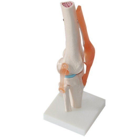
Human Knee Joint Teaching Model Training Model
Approx $62.08 USD
Introduction to the Human Knee Joint Teaching Model
The Human Knee Joint Teaching Model is an essential educational tool designed to support in-depth study of knee anatomy and function. This model provides a realistic representation of the knee joint, allowing students, educators, healthcare professionals, and trainers to gain a better understanding of its structure and mechanics. With detailed features like ligaments, cartilage, and bone surfaces, this model serves as an invaluable aid for learning about knee injuries, treatment options, and rehabilitation practices. Ideal for use in medical classrooms, clinics, and physical therapy training programs across New Zealand, this model enhances learning by offering a tangible, interactive way to explore knee anatomy.
H2: Key Features of the Human Knee Joint Teaching Model
1. Accurate Representation of Knee Anatomy
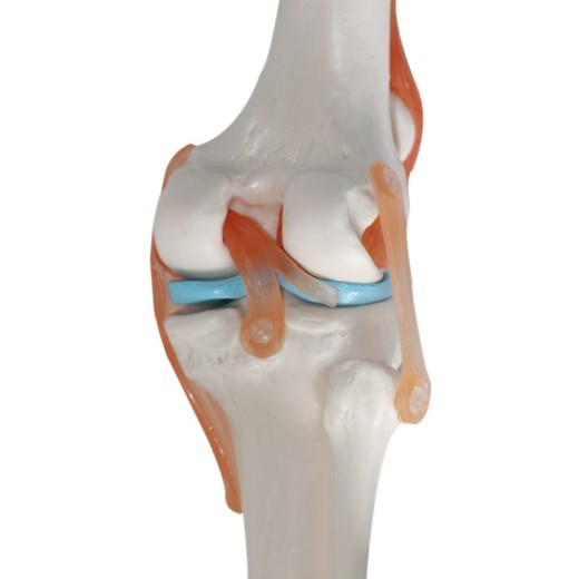
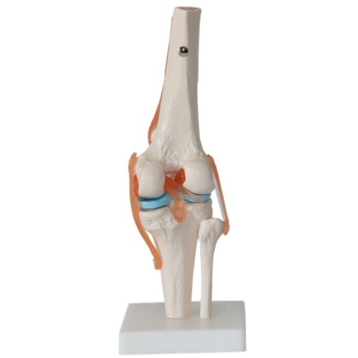
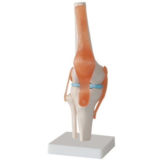
The Human Knee Joint Model provides a realistic depiction of the major components of the knee joint, including the femur, tibia, fibula, patella, and key ligaments (ACL, PCL, MCL, and LCL). Each part is precisely crafted to mimic the anatomical structure of a real knee, allowing users to visualize the complexity of the joint and understand how these components work together to provide stability and movement.
2. High-Quality and Durable Materials
Constructed from high-grade, non-toxic materials, this model is built to withstand frequent use in educational and clinical settings. The durable construction ensures it remains intact even after extensive handling, making it suitable for classrooms, orthopedic clinics, and physiotherapy training centers. This long-lasting quality makes it a reliable resource for ongoing education, providing excellent value over time.
3. Functional Movement to Demonstrate Joint Mechanics
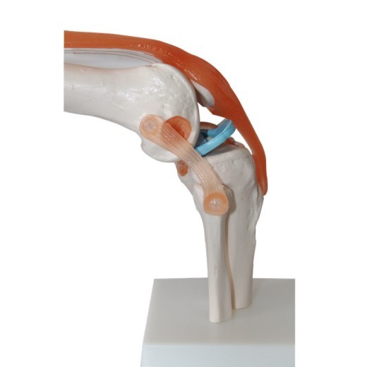
The knee model features movable parts that replicate the natural movements of the knee joint, including flexion, extension, and slight rotation. This functionality allows students and healthcare professionals to observe how the knee moves, helping to explain joint mechanics, muscle action, and the role of each ligament. By simulating real joint motion, this model provides a dynamic learning experience that enhances comprehension of knee functionality and common issues, such as ligament tears and arthritis.
4. Color-Coded Parts for Easy Identification
To aid in learning, the model includes color-coded ligaments, cartilage, and bone structures, allowing users to easily identify each part. The color coding helps beginners quickly distinguish between different structures, making it easier to understand the roles and relationships of each component within the knee. This feature is especially beneficial for visual learners, as it enhances memorization and understanding of the anatomy.
5. Compact and Display-Friendly Design
The compact design of the knee joint model makes it easy to display on desks, shelves, or in medical classrooms without taking up excessive space. It comes with a sturdy base that keeps the model stable during demonstrations and can be easily moved between different teaching or consultation areas. This portability makes it suitable for a variety of settings, whether used as part of a classroom demonstration or displayed in a clinic for patient education.
6. Ideal for Clinical Training and Patient Education
The Human Knee Joint Model is not only useful for students but also serves as an excellent tool for patient education in clinics. Orthopedic specialists, physical therapists, and sports medicine professionals can use this model to explain knee injuries, treatment options, and rehabilitation steps to patients, providing a clear and understandable visual reference. This aids in patient understanding and fosters better communication between healthcare providers and their patients.
H2: Why Choose the Human Knee Joint Teaching Model?
1. Essential for Medical and Health Education
The knee joint model is a vital resource for teaching the anatomy and function of one of the body’s most important joints. For students in medical and health-related fields, it provides a reliable way to learn the complexities of knee anatomy and to understand common issues like ligament injuries, cartilage wear, and joint degeneration. This hands-on model makes it easier to connect theoretical knowledge with real-world applications, enhancing the educational experience.
2. Suitable for Professional Training Programs
In professional training settings, such as orthopedic residencies, physical therapy courses, and sports medicine workshops, this model is invaluable for understanding knee mechanics and common injuries. Trainers can use the model to illustrate the mechanisms behind ACL tears, patellar dislocations, and other injuries, providing an effective way to study diagnosis, treatment, and rehabilitation techniques. For New Zealand’s healthcare education programs, this model is a key tool for advancing anatomical knowledge and clinical skills.
3. Interactive Tool for Visual and Kinesthetic Learners
The Human Knee Joint Teaching Model caters to both visual and kinesthetic learners, making it a versatile tool for different learning styles. The color-coded parts support visual learning by making it easy to identify each component, while the movable parts allow kinesthetic learners to physically engage with the model. This interactive approach enhances comprehension, helping learners of all types retain information about knee structure and function.
4. Perfect for Exam Preparation and Practical Assessments
Students preparing for exams or practical assessments will find this model helpful for reviewing knee anatomy and mechanics. Its realistic representation and labeled parts make it an effective study aid, allowing students to reinforce their understanding of key structures and joint functions. For medical students, physiotherapists, and orthopedic trainees, the Human Knee Joint Model serves as a practical tool that builds confidence and knowledge.
5. Educational Display for Clinics and Classrooms
Beyond its educational function, the Human Knee Joint Model is an excellent display piece for clinics, classrooms, and rehabilitation centers. It serves as a visual reference for patients and students, providing insight into knee structure and the impact of various injuries. For New Zealand educators and healthcare providers, the model offers an effective way to foster a greater understanding of knee anatomy and encourage conversations about joint health.
H2: Maintenance and Care Tips for Your Human Knee Joint Teaching Model
To keep your Human Knee Joint Teaching Model in top condition, follow these maintenance tips:
-
Regular Cleaning: Use a soft, dry cloth to dust the model frequently, especially around the moving parts and color-coded
sections. For deeper cleaning, a damp cloth with mild soap is sufficient. Avoid harsh chemicals to prevent damage.
-
Handle with Care: Though durable, the model’s moving parts should be handled gently to prevent wear or misalignment. Avoid
forcing any part beyond its intended range of motion to ensure it remains functional.
-
Store Safely: When not in use, store the model in a safe, dry area away from direct sunlight. Proper storage helps protect
it from dust, potential damage, and fading of color-coded parts.
- Avoid Excessive Sunlight: Prolonged exposure to sunlight can fade colors. Place the model in shaded or indoor areas to maintain its appearance and ensure longevity.
Product information:
Size: naturally large, fixed on the substrate.
Material: PVC material
Size: 17x14.5x29.5cm
Packing list:
Plastic joint model * 1
Product Image:


The product may be provided by a different brand of comparable quality.
The actual product may vary slightly from the image shown.



.webp)





.webp)


.webp)



.jpg)


.jpg)


.jpeg)


.jpeg)


.jpeg)


.jpeg)


.jpeg)

.jpeg)

.jpeg)

.jpeg)


.jpeg)
.jpg)

.jpeg)


.jpeg)
.jpeg)


.jpeg)


.jpeg)


.jpeg)

.jpeg)

.jpeg)

.jpeg)


.jpeg)
.jpeg)
.jpeg)


.jpeg)


.jpeg)




.jpg)
.jpeg)


.jpg)


ulva-Logo.jpg)


.jpeg)


.png)


.png)


