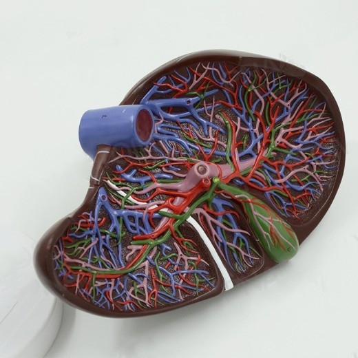
Liver Anatomical Model Hepatic Portal Hepatic Lobe Model
Approx $69.84 USD
Introduction to the Liver Anatomical Model Hepatic Portal and Hepatic Lobe Model
The Liver Anatomical Model with Hepatic Portal and Hepatic Lobe details is an advanced educational tool designed to support in-depth learning of liver anatomy and function. This model provides a realistic view of the liver’s structure, including the hepatic portal system and distinct hepatic lobes. Ideal for students, educators, and healthcare professionals, this model is valuable in classrooms, clinics, and anatomy labs across New Zealand, offering a hands-on, three-dimensional approach to studying liver health, blood flow, and digestive functions. With detailed visualization of both the hepatic portal system and lobular structure, this model is essential for understanding the complexities of liver anatomy.
H2: Key Features of the Liver Anatomical Model Hepatic Portal and Hepatic Lobe Model
1. Detailed Representation of the Liver and Hepatic Portal System
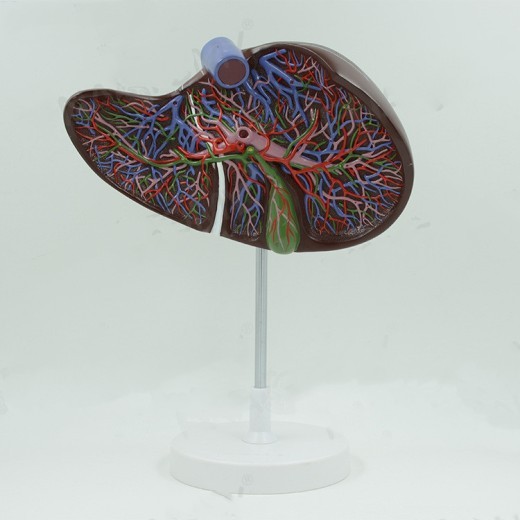
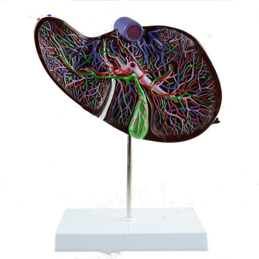
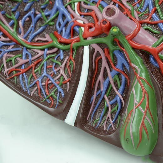
The Liver Anatomical Model accurately showcases major anatomical features, including the hepatic portal vein, hepatic arteries, and bile ducts. It allows users to visualize how blood flows into and out of the liver, making it easier to understand processes like detoxification, metabolism, and nutrient processing. This level of detail is invaluable for students and professionals who need a thorough understanding of liver function.
2. Clear Division of Hepatic Lobes
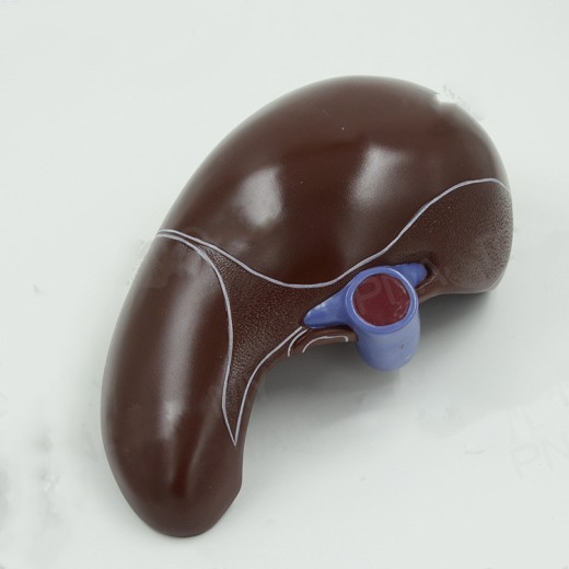
The model clearly delineates the four primary lobes of the liver: the right, left, caudate, and quadrate lobes. Each lobe is precisely represented, helping users learn the structural organization and functional segments within the liver. This feature is especially useful for medical students and healthcare providers who need to identify specific liver regions in clinical contexts.
3. High-Quality and Durable Construction
Constructed from high-grade, non-toxic materials, the model is designed to withstand frequent handling, making it suitable for educational and clinical settings. Its durable build ensures longevity, providing a reliable resource for anatomy classrooms, medical labs, and healthcare training centers. This quality construction means it can be used repeatedly over time, delivering excellent value to New Zealand’s educational institutions.
4. Color-Coded and Labeled Parts for Easy Identification
The Liver Anatomical Model includes color-coded sections to help users quickly differentiate between hepatic structures, blood vessels, and ducts. This color-coding makes it easier to identify each part of the liver, such as the hepatic lobes, bile ducts, and hepatic portal vein. Labels provide clear guidance, which is especially useful for beginners and visual learners, enhancing comprehension and memorization of liver anatomy.
5. Cross-Sectional View for Enhanced Study
The model often features a cross-sectional design that allows users to see the inner structure of the liver, including lobular and vascular details. This cross-sectional view offers insight into how the hepatic lobes connect and how blood flows within the liver, supporting a better understanding of complex processes such as filtration, bile production, and metabolism. This feature makes the model especially valuable for advanced studies in hepatology and internal medicine.
6. Compact and Display-Ready Design
Designed to be compact and portable, this liver model can be displayed on desks, shelves, or demonstration areas without taking up too much space. Its stable base keeps the model secure during demonstrations and discussions, making it easy to move between classrooms, labs, and consultation rooms. This display-ready feature is ideal for a variety of settings, allowing educators and healthcare professionals in New Zealand to use it flexibly.
H2: Why Choose the Liver Anatomical Model Hepatic Portal and Hepatic Lobe Model?
1. Essential for Medical and Health Education
The Liver Anatomical Model is an indispensable resource for teaching liver anatomy in medical and health sciences. For students and trainees, it offers a clear view of liver structure and function, bridging the gap between theoretical knowledge and practical understanding. This model makes anatomy lessons more interactive and accessible, providing a deeper comprehension of how the liver supports vital body functions.
2. Perfect for Hepatology and Gastroenterology Training
This liver model is invaluable for training in hepatology, gastroenterology, and internal medicine. Trainers can use it to illustrate the structure and function of the hepatic portal system, bile production, and liver lobular organization. For New Zealand’s healthcare training programs, this model offers a practical, hands-on tool for understanding liver health and pathology, from liver diseases to hepatic surgeries.
3. Supports Visual and Kinesthetic Learning
This model is beneficial for visual and kinesthetic learners, offering a hands-on experience that enhances the understanding of liver anatomy. The color-coded parts support visual learning, while the cross-sectional features allow kinesthetic learners to explore internal structures directly. This interactive approach makes it easier to retain information and grasp the liver’s complexity.
4. Ideal for Exam Preparation and Practical Learning
For students preparing for exams or practical assessments, the Liver Anatomical Model serves as a valuable study aid. Its realistic design and labeled parts make it easy to review key structures and liver functions, reinforcing anatomical knowledge. Medical students and trainees can use this model to build confidence and enhance their understanding before assessments, helping them perform better in both academic and clinical environments.
5. Educational Display for Clinics and Classrooms
Beyond its educational value, this model serves as a useful display piece in clinics, classrooms, and laboratories. In clinical settings, it can be used to explain liver diseases, surgical procedures, and treatment options to patients, improving their understanding of liver health. For New Zealand educators and healthcare providers, this model provides an effective way to foster greater awareness and understanding of the liver’s role in health and disease.
H2: Maintenance and Care Tips for Your Liver Anatomical Model with Hepatic Portal and Hepatic Lobe Model
To keep your Liver Anatomical Model in excellent condition, follow these care tips:
-
Dust Regularly: Use a soft cloth or brush to dust the model, especially around the color-coded sections and detailed areas.
Regular cleaning preserves the model’s appearance and prevents dirt buildup.
-
Avoid Direct Sunlight: Display the model away from direct sunlight to prevent colors from fading over time. Indoor
placement helps maintain its vibrant appearance and quality.
-
Handle with Care: While durable, handle the model gently to avoid damaging delicate parts. Avoid applying excessive
pressure, especially around smaller, intricate structures like the hepatic portal vein and bile ducts.
- Store Properly When Not in Use: When not on display, store the model in its original packaging or a protective case to prevent dust accumulation and potential damage. Proper storage helps extend the model’s lifespan for long-term educational use.
Product Information
Liver Model Liver Model With Blood Vessels, Liver Anatomical Model Liver And Gallbladder Enlargement Model Liver And Gallbladder Model
This Model Shows The Lobes Of The Liver, The Vascular Branches Of The Liver, The Gallbladder, And The Bile Duct System. Different Colors Indicate The Complete Vascular Network In The Liver: Portal Vessels, Intrahepatic And Extrahepatic Bile Ducts, Placed On A Detachable Base Frame. . 1.5 Times Magnification, Made Of Imported Pvc Material, Hand-Painted, Very Beautiful


The product may be provided by a different brand of comparable quality.
The actual product may vary slightly from the image shown.


.webp)
.webp)





.jpg)


.jpg)


.jpeg)


.jpeg)


.jpeg)


.jpeg)


.jpeg)

.jpeg)

.jpeg)

.jpeg)


.jpeg)
.jpg)

.jpeg)


.jpeg)
.jpeg)


.jpeg)


.jpeg)


.jpeg)

.jpeg)

.jpeg)

.jpeg)


.jpeg)
.jpeg)
.jpeg)


.jpeg)


.jpeg)


.jpg)
.jpeg)


.jpg)


ulva-Logo.jpg)


.jpeg)


.png)


.png)


