Mini Skull Anatomical Model Can Disassemble The Skull
Approx $53.21 USD
Introduction to the Mini Disassemblable Skull Anatomical Model
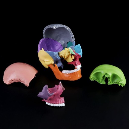
The Mini Disassemblable Skull Anatomical Model is a compact, realistic representation of the human skull, designed to provide a detailed look at cranial anatomy. With detachable parts, this model allows students, healthcare professionals, and educators to explore the individual bones and structures of the skull. Ideal for anatomy classrooms, medical training centers, and clinics across New Zealand, this model is perfect for studying skull composition, cranial sutures, and bone alignment in a hands-on way. Its mini size and disassemblable design make it convenient for close examination and interactive learning.
H2: Key Features of the Mini Disassemblable Skull Anatomical Model
1. Realistic and Detailed Skull Anatomy
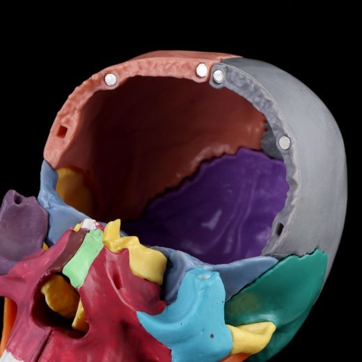
This model provides a realistic depiction of the human skull, including essential features like cranial bones, jaw structure, eye sockets, nasal cavity, and cranial sutures. The detailing allows users to study individual bones in the skull and observe how they fit together to form a protective structure for the brain. This feature is ideal for those interested in the intricacies of human cranial anatomy.
2. Detachable Parts for Interactive Learning
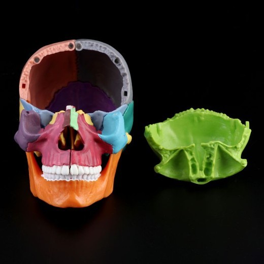
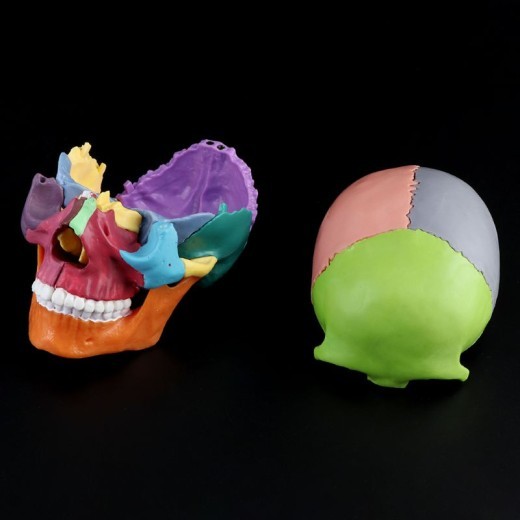
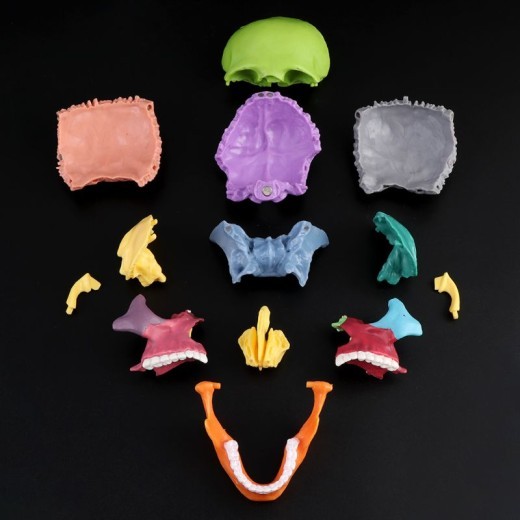
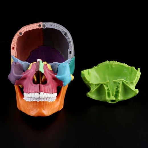
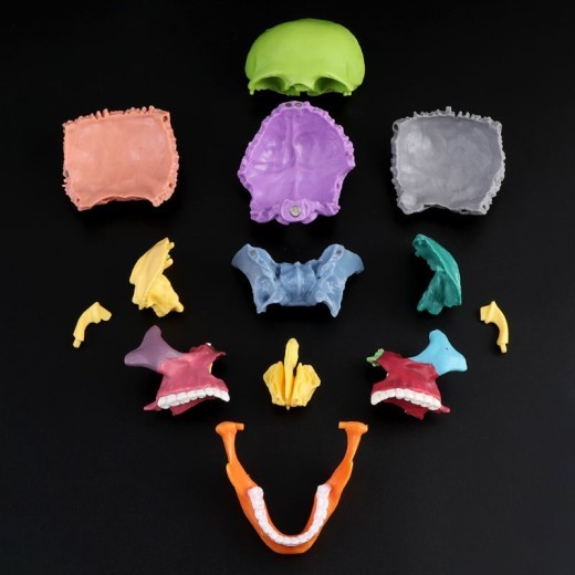
The mini skull model is designed to disassemble into multiple parts, including the skull cap, mandible (lower jaw), and other key sections. This disassemblable feature allows users to study each bone separately and understand how they interconnect, enhancing comprehension of cranial structure and function. It’s especially valuable for hands-on learners who benefit from exploring physical models as part of the educational experience.
3. Compact and Portable Design
Standing at a convenient mini size, this model is easy to transport and display on desks, tables, or shelves. Its compact design makes it suitable for individual study, classroom demonstrations, or on-the-go learning. This portability is beneficial for educators and students in New Zealand who require a space-efficient and mobile anatomy resource.
4. Color-Coded Bones for Easy Identification
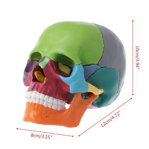
Many mini skull models come with color-coded bones, making it easier to identify and differentiate between various cranial structures, such as the frontal, parietal, temporal, and occipital bones. This color-coding aids visual learners and makes the learning process more intuitive, allowing users to quickly recognize each cranial section.
5. High-Quality and Durable Materials
Constructed from non-toxic, high-quality materials, this model is designed to withstand regular handling in educational and clinical environments. Its durable build ensures it retains detail over time, making it ideal for frequent use in anatomy labs, classrooms, and medical facilities. The high-quality materials make the model a long-lasting resource for anatomy study in New Zealand.
6. Stable Base for Display
The model typically includes a stable base to keep it secure and upright, allowing for easy viewing and interaction. This stability is especially useful during demonstrations or study sessions, where users may need to handle and examine various parts of the skull without worrying about the model tipping over.
H2: Why Choose the Mini Disassemblable Skull Anatomical Model?
1. Essential for Anatomy and Medical Education
This mini skull model is an invaluable tool for teaching cranial anatomy, particularly for students in medical and health sciences programs. By offering a clear and interactive view of the skull’s structure, it helps learners understand the spatial relationships between different cranial bones. For students and professionals in New Zealand, this model bridges the gap between theoretical learning and hands-on exploration.
2. Perfect for Exam Preparation and Practical Assessments
For students preparing for exams or practical assessments, this model serves as a valuable study aid. The detachable parts allow users to review and memorize key bones and sutures, reinforcing knowledge that’s crucial for anatomy exams. Medical students and healthcare trainees benefit from this interactive approach to studying cranial anatomy, enhancing retention and confidence.
3. Supports Visual and Kinesthetic Learning
The model caters to both visual and kinesthetic learners, offering a hands-on experience that enhances understanding. Color-coded sections support visual learning by distinguishing between different bones, while the disassemblable parts allow kinesthetic learners to interact directly with the model. This interactivity makes learning about cranial anatomy more effective and engaging.
4. Ideal for Patient Education in Clinical Settings
In clinical settings, this mini skull model can help healthcare providers explain cranial structure, injuries, or conditions related to the head and neck. By allowing patients to see and touch a model of the skull, providers can make anatomy more accessible, improving patient understanding and communication.
5. Educational Display for Classrooms and Clinics
Beyond its educational value, the Mini Skull Anatomical Model serves as an informative display for classrooms, clinics, and health education centers. In educational settings, it provides a visual aid for explaining cranial anatomy, supporting discussions on head protection and brain health. For educators and healthcare providers in New Zealand, this model enhances anatomical understanding in a visually compelling way.
H2: Maintenance and Care Tips for Your Mini Disassemblable Skull Anatomical Model
To keep your model in excellent condition, follow these care tips:
-
Dust Regularly: Use a soft cloth or brush to remove dust from the model, particularly around intricate areas like sutures
and joints. Regular cleaning helps maintain the model’s appearance and prevents dirt buildup.
-
Avoid Direct Sunlight: Display the model out of direct sunlight to prevent colors from fading over time, preserving the
model’s vibrant look and quality.
-
Handle with Care: Although durable, handle the detachable parts gently to avoid wear or damage, especially when separating
or reassembling smaller bones.
- Store Properly When Not in Use: When not displayed, store the model in its original packaging or a dust-free area to protect it from potential damage. Proper storage helps extend the model’s lifespan for long-term educational use.
Product Details:
Color assembled mini skull, 15 parts Product size: 22.8*6.8*29.5cm
product description:
These two models are human internal organ models composed of 15 parts. You need to assemble these parts yourself into a complete human
internal organ model. By assembling this model, it is helpful to understand the composition and structure of human organs, and it is also
very suitable for preschool education system as a teaching use. In the assembling process, the coordination of the left and right hands and
the coordination of the hand and the eye are required to exercise the flexibility and coordination of the fingers and the coordination
between the hands and the eyes. More importantly, it develops the potential of the space imagination and the right brain. After assembling
these products, you can enjoy the joy of success and bring a sense of accomplishment and satisfaction.


The product may be provided by a different brand of comparable quality.
The actual product may vary slightly from the image shown.





.webp)
.webp)
.webp)
.webp)










.jpg)


.jpg)


.jpeg)


.jpeg)


.jpeg)


.jpeg)


.jpeg)

.jpeg)

.jpeg)

.jpeg)



.jpeg)
.jpg)

.jpeg)


.jpeg)
.jpeg)


.jpeg)


.jpeg)


.jpeg)

.jpeg)

.jpeg)

.jpeg)


.jpeg)
.jpeg)
.jpeg)


.jpeg)


.jpeg)




.jpg)
.jpeg)


.jpg)


ulva-Logo.jpg)


.jpeg)


.png)













.png)
